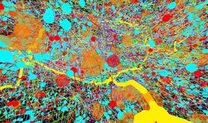
Reconstruction of part of the nervous system in a fruit fly (Drosophila melanogaster) larva. Neural circuits were digitally reconstructed from serial section transmission electron micrographs through an abdominal segment of the nervous system. This image shows a lateral view of neurones and neuronal synapses in the neuropil (area in the grey matter of the central nervous system where neurones form connections with each other) of a segment of the nerve cord. A single axon from a sensory neurone that senses vibration (a chordotonal) is visible in yellow. Each coloured line belongs to the skeletonised representation of a neuronal branch (arbor). The points of contact between two neurones, the synapses, are shown in solid blue (input side) and solid red (output side). The transparent orange spheres represent labelled points of interest on the skeletons of the neurones, such as the presence of mitochondria and other features. Horizontal width of image is approximately 15 micrometres. Wellcome Image Awards 2015.