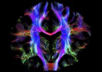
Coronal view of nerve fibres in the brain of a young healthy adult, which has been virtually sliced down a vertical axis to divide it into front and back. The brain is viewed from behind, with the left side of the brain on the left of the image. This image was created by virtually dissecting the brain using data obtained from diffusion magnetic resonance imaging (MRI). Diffusion weighted imaging is a specialised type of MRI scan which measures water diffusion in many directions in order to reconstruct the orientation of bundles of nerve fibres. Tractography is used to indirectly model these nerve fibres, which transmit information between different regions of the brain. These have been colour-coded to help distinguish between different tracts which pass close to each other. For example, fibres connecting the left and right hemispheres (red), fibres travelling from top to bottom (blue) connecting to the spinal cord, and fibres running from front to back (green) are visible here. Reconstructing these connections between different parts of the brain will aid our understanding of how the brain functions in health and disease, and could ultimately become a tool in the same way as the human genome. Width of image is approximately 165 mm. Magnetic resonance imaging 2015.