Thigh bone (femur) from a male Japanese quail, micro-CT

  • Justyna Miszkiewicz, Jayashree Chakraborty, John Logan, Duncan Bassett, Graham Williams, Imperial College London
  • Digital Images
  • Online

Available online

view Thigh bone (femur) from a male Japanese quail, micro-CT

Attribution-NonCommercial 4.0 International (CC BY-NC 4.0)

You can use this work for any purpose, as long as it is not primarily intended for or directed to commercial advantage or monetary compensation. You should also provide attribution to the original work, source and licence. Read more about this licence.

Credit

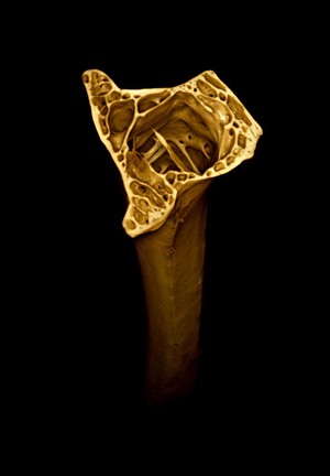
Thigh bone (femur) from a male Japanese quail, micro-CT. Justyna Miszkiewicz, Jayashree Chakraborty, John Logan, Duncan Bassett, Graham Williams, Imperial College London. Attribution-NonCommercial 4.0 International (CC BY-NC 4.0). Source: Wellcome Collection.

Selected images from this work

View 1 image

About this work

Description

3D reconstruction of high-resolution, micro-computer tomography (CT) scans of the thigh bone (femur) from a male Japanese quail (Coturnix japonica). The delicate and intricate trabecular bone structure within the marrow cavity of the femur is shown here. The medullary bone used to store calcium for eggshell formation in female quails is absent from the marrow cavity in males. Average length of an intact quail femur is approximately 40 mm. Like many other birds, quails control the timing of their reproduction through a physiological process called photoperiodism. Quails are highly sensitive to day length and use this information to ensure that reproduction occurs during the most favourable season for their offspring (i.e. in the summer). Increase in day length results in a series of hormonal changes leading to greater oestrogen production and fertility. In birds, these hormonal changes also result in the formation of extensive "medullary bone" within the marrow cavity. This medullary bone is essential to supply the large amount of calcium required to form the eggshell.

Permanent link